
Pathology scoring plays a central role in biomedical research, drug development, biomarker evaluation, and disease characterization. Whether researchers are grading tumor morphology, quantifying immunohistochemical staining, assessing fibrosis, measuring inflammation, or evaluating treatment-induced tissue changes, histopathological scores often become important endpoints for comparing experimental groups and patient cohorts.
However, conventional pathology scoring has an unavoidable challenge: inter-reader variability.
Two experienced pathologists examining the same slide can sometimes assign different scores. Even the same reader may interpret borderline cases differently at separate time points. When hundreds or thousands of tissue samples are distributed across study sites, laboratories, scanners, staining batches, and cohorts, these small differences can accumulate into a major reproducibility problem.
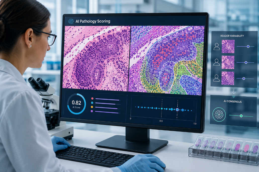
This is where pathology scoring AI is becoming increasingly valuable. Instead of replacing pathological expertise, artificial intelligence can provide a consistent computational reference that applies the same image-analysis rules across every slide. By combining whole-slide imaging, deep learning, automated segmentation, feature quantification, and standardized scoring algorithms, AI can help researchers reduce reader-dependent variation and generate more comparable pathology data across cohorts.
Why Is Inter-Reader Variability a Persistent Problem in Pathology?
Histopathology is fundamentally an interpretation of complex biological patterns. Unlike a laboratory measurement that produces a single numerical output, tissue assessment frequently requires a pathologist to integrate information about cellular morphology, staining intensity, tissue architecture, lesion distribution, and disease-specific criteria.
This creates several potential sources of variability.
Different Interpretations of Scoring Criteria
Many histopathological endpoints use categorical or semi-quantitative scoring systems. A pathologist may need to determine whether a feature should receive a score of 1, 2, or 3 based on its severity or extent.
The problem becomes particularly obvious around category boundaries. One reader may interpret a tissue feature as mild-to-moderate and assign a lower category, while another reader considers the same feature sufficiently extensive to justify the next category.
These differences do not necessarily mean that one reader is wrong. They reflect the inherent difficulty of translating continuous biological changes into discrete scoring categories.
Variation in Region Selection
A whole-slide image can contain millions or even billions of pixels and extensive areas of heterogeneous tissue. Manual assessment frequently depends on which regions the reader chooses to examine most closely.
This is especially relevant for heterogeneous biomarkers and lesions.
For example, a biomarker may be strongly expressed in one tumor region but weakly expressed elsewhere. Fibrosis may form focal deposits rather than being evenly distributed throughout the tissue. Mitotic figures can also concentrate in specific hotspots.
If readers select different fields of view or regions of interest, their final scores can diverge even when they apply similar scoring rules.
Subjective Estimation of Percentages
Pathologists are often asked to estimate percentages, such as the fraction of positive tumor cells, percentage of fibrotic tissue, proportion of necrosis, or percentage of cells showing a particular staining intensity.
Human visual estimation is highly valuable, but it is not equivalent to counting every cell or measuring every relevant pixel.
This becomes especially important near clinically or experimentally meaningful cutoffs. A difference between an estimated 8% and 12%, for example, may appear small quantitatively but could move a sample into a different predefined category.
Studies using AI-assisted pathology illustrate this problem. In a Swiss national study of tumor cell fraction assessment, computer-aided support reduced the standard deviation of pathologists’ estimates relative to the reference from 9.9% to 5.8%, while the intraclass correlation coefficient increased from 0.80 to 0.93.
Differences Between Laboratories and Cohorts
Inter-reader variability becomes even more complicated in multi-cohort research.
Samples may differ in:
- tissue fixation and processing;
- section thickness;
- staining protocols;
- reagent batches;
- slide scanners;
- image resolution;
- color profiles;
- disease stage;
- specimen type;
- tissue composition.
These differences introduce what is often called domain variation or domain shift in digital pathology. A scoring method that performs well on slides generated in one laboratory cannot automatically be assumed to behave identically on slides produced elsewhere.
This issue affects both human readers and AI systems. Research on AI-based prostate cancer grading, for example, has shown that differences in sample preparation, including staining and sectioning variables, can influence model performance, emphasizing the importance of robust external validation across heterogeneous datasets.
How Pathology Scoring AI Creates a More Consistent Reference
The key advantage of AI is not simply speed. Its greater value for cohort-level pathology studies is repeatability.
Once an algorithm and its parameters are fixed, the same computational rules can be applied systematically across a dataset.
1. Standardized Tissue and Cell Detection
Modern pathology AI models can segment tissue regions and identify structures such as tumor cells, immune cells, nuclei, glands, fibrotic areas, necrosis, or other disease-related features.
Instead of asking each reader to subjectively decide where a structure begins or ends, an appropriately validated AI pipeline uses a consistent segmentation process.
This can be particularly useful when a study contains thousands of images and consistent annotation at scale would otherwise be difficult.
2. Consistent Quantification
After detecting relevant structures, AI can convert visual patterns into numerical variables.
Depending on the study, these may include:
- positive cell percentage;
- cell density;
- staining intensity;
- tumor-to-stroma ratio;
- fibrosis area;
- lesion burden;
- immune-cell infiltration;
- mitotic count;
- necrotic area;
- biomarker-positive area.
Quantitative measurements make it easier to compare samples without relying exclusively on broad visual categories.
Rather than describing two tissue samples simply as “moderate,” researchers can potentially compare their measured biological features on a continuous scale. Categorical scores can then be generated according to predefined rules when required.
3. Reproducible Threshold Application
Cutoff-dependent scoring is one of the areas in which inter-reader variability can have the greatest practical impact.
An algorithm can apply the same threshold to every eligible cell or region. This provides a stable reference when human assessment is difficult, particularly for borderline samples.
PD-L1 scoring provides an informative example. A multicenter study of urothelial carcinoma used an AI-powered combined positive score analyzer on cases collected from three institutions. Before AI-guided reevaluation, complete agreement among three pathologists was observed in 82.1% of cases. Following AI-guided review of discrepant cases, complete agreement increased to 93.9%, and variability associated with the source hospital was reduced.
The point is not that AI should automatically determine every final score. Rather, a computational score can function as a standardized reference point when readers disagree.
From Single Slides to Cross-Cohort Standardization
The real importance of pathology scoring AI becomes apparent when datasets expand beyond a single laboratory.
Consider a preclinical study involving several treatment groups evaluated at different time points. Alternatively, imagine a multicenter translational study in which tissue is obtained from multiple institutions.
Without harmonization, apparent biological differences between cohorts may partly reflect differences in slide preparation or interpretation.
AI can support cohort standardization at several levels.
A Common Analytical Pipeline
All images can pass through the same computational workflow:
image quality control → tissue detection → region segmentation → feature extraction → quantitative measurement → scoring → data export
Applying one predefined pipeline reduces methodological differences introduced when readers or sites independently develop their own interpretation habits.
Continuous Measurements Instead of Only Categories
Another important advantage is the ability to retain quantitative information underneath conventional grades.
Suppose fibrosis is traditionally reported using stages 0 through 4. Two samples categorized as stage 2 may nevertheless have meaningfully different fibrosis distributions.
An AI system can potentially record quantitative tissue features while still producing a conventional stage.
This creates a richer dataset for downstream analyses, including treatment-response studies, longitudinal disease monitoring, biomarker discovery, and predictive modeling.
AI-assisted fibrosis assessment is already being investigated in metabolic dysfunction-associated steatohepatitis. In a randomized crossover study involving 120 digitized slides from two trials and four expert hepatopathologists, AI assistance improved inter-pathologist agreement in fibrosis staging, particularly in earlier-stage fibrosis.
Consistent Reanalysis of Historical Data
AI also makes it possible to apply a newly established analytical definition retrospectively.
If a scoring protocol changes during a long research program, manually rereading thousands of archived slides can require considerable time. With digitized slides and an appropriate validated model, the entire dataset can potentially be reprocessed through the same version of an algorithm.
This makes longitudinal comparisons easier and provides clearer version control over how measurements were generated.

AI as a Second Reader Rather Than a Replacement
A useful way to think about pathology AI is as a consistent computational second reader.
Pathologists bring biological understanding, disease context, recognition of unusual morphology, and the ability to identify situations that fall outside a model’s intended scope. AI brings repeatability, large-scale quantification, and the ability to apply identical numerical rules across images.
Combining these strengths can improve scoring consistency.
For instance, AI-assisted Gleason grading has been evaluated in prostate biopsy assessment. In a study involving 14 observers, AI assistance significantly increased agreement between readers and an expert reference standard.
More recently, research into AI-supported HER2 scoring has similarly demonstrated the potential of computational assistance to improve agreement among human readers. A study involving 853 HER2 immunohistochemistry whole-slide images reported an increase in overall multireader agreement from 79.8% without AI support to 88.6% with AI assistance.
These findings illustrate an important point: the strongest workflow is often not human versus AI, but human plus AI.
What Makes an AI Scoring System Reliable Across Cohorts?
Consistency should not be confused with accuracy.
An algorithm can consistently produce the same result and still be systematically wrong. Therefore, reducing inter-reader variability requires more than simply deploying a deep-learning model.
Representative Training Data
Training data should capture the variation the model will encounter in real-world studies. This may include different staining intensities, tissue types, disease stages, scanners, laboratories, and sample qualities.
A narrowly trained model may perform well internally but deteriorate when applied to a new cohort.
Independent Validation
Validation should ideally include samples that were not used for model training.
For multicenter applications, external cohorts are particularly important because they help determine whether the algorithm remains reliable when laboratory and patient characteristics change.
Image Quality Control
Poorly focused slides, tissue folds, staining artifacts, bubbles, pen marks, damaged sections, and scanning errors can affect computational measurements.
Automated or semi-automated quality control should therefore be incorporated before scoring.
A model should also be able to flag images that fall outside acceptable quality thresholds rather than silently generating potentially misleading measurements.
Version Control
AI models can evolve.
Retraining a model, changing segmentation parameters, modifying a threshold, or updating preprocessing steps may change output values. Researchers should therefore record which model version generated each dataset.
For longitudinal programs, controlled model versioning is essential for maintaining comparability.
Human Review of Discordant Cases
The most informative cases are often those where the AI and the pathologist disagree.
Instead of treating disagreement as a failure, these samples can be reviewed to identify:
- unusual tissue morphology;
- staining artifacts;
- segmentation errors;
- borderline biological patterns;
- ambiguous scoring criteria;
- previously underrepresented tissue features.
This feedback can improve both human scoring guidelines and future versions of the computational model.
Why Reduced Inter-Reader Variability Matters for Preclinical Research
In preclinical studies, pathology endpoints are frequently used to determine whether a therapeutic candidate changes disease severity or produces tissue toxicity.
When treatment effects are subtle, measurement variability can obscure biological signals.
A standardized pathology scoring AI workflow can help make group-level comparisons more reliable by ensuring that control and treatment samples are analyzed according to the same computational criteria.
This is particularly valuable for longitudinal or large-scale studies involving:
- efficacy evaluation;
- toxicologic pathology;
- fibrosis research;
- oncology models;
- inflammatory disease models;
- biomarker studies;
- organ-specific disease models.
The objective is not simply to generate a score faster. It is to make the score more comparable across animals, experimental groups, study phases, readers, sites, and cohorts.
Moving From Subjective Scoring Toward Quantitative Pathology
Traditional categorical scoring systems remain highly useful because they summarize complex morphology in a form that researchers and clinicians understand.
AI does not necessarily eliminate these scoring systems. Instead, it can add a quantitative layer underneath them.
A future pathology report might therefore contain both:
Conventional assessment: Fibrosis stage = 2
AI-derived measurements: Fibrotic area percentage, collagen distribution, regional density, morphological pattern metrics, and confidence indicators
This combination preserves an interpretable biological category while providing continuous variables suitable for statistical analysis and predictive modeling.
As digital pathology datasets become larger and increasingly integrated with molecular, imaging, and preclinical data, this quantitative approach may also improve connections between tissue morphology and downstream computational disease models.
Conclusion: Standardization Is the Real Value of Pathology AI
Inter-reader variability is not simply a problem of individual expertise. It results from the inherent complexity of tissue morphology, subjective thresholds, heterogeneous samples, regional selection, and differences between laboratories and cohorts.
Pathology scoring AI addresses this problem by creating a repeatable computational framework.
The same model can identify structures, measure features, apply thresholds, and calculate scores according to predefined criteria across large numbers of whole-slide images. When combined with expert pathological review, this approach can reduce inter-reader variability, improve reproducibility, identify discordant cases, and make data from different study cohorts easier to compare.
For research organizations working with increasingly large digital pathology datasets, the most meaningful transition is therefore not simply from manual pathology to automated pathology. It is the transition from subjective visual estimates alone toward standardized, quantitative, and human-supervised pathology assessment.
Related AI Services from Creative Biolabs
Creative Biolabs provides several AI-driven services that can support pathology and preclinical research:
- AI-Driven Pathology Image Analysis Service – Automated analysis of digital pathology images for feature detection, quantification, classification, and standardized assessment.
- AI-Driven Preclinical Research Service – AI-supported analysis and predictive solutions for preclinical drug research and development.
- AI-Driven Disease Model Construction & Prediction Service – AI-based disease modeling and prediction for disease progression, therapeutic response, and preclinical research.
Explore these services to learn how Creative Biolabs can support your AI-driven pathology and preclinical research projects.