Whole-slide imaging (WSI) has changed how pathology data can be captured, stored, reviewed, and analyzed. In preclinical research, this shift is especially important because tissue slides are often used to evaluate disease progression, treatment response, organ toxicity, immune infiltration, fibrosis, necrosis, inflammation, and other biological changes that directly influence candidate selection. As artificial intelligence becomes more integrated into digital pathology, researchers are no longer limited to qualitative descriptions or semi-quantitative scoring. Instead, AI pathology image analysis can help transform WSI datasets into structured, reproducible, and statistically usable endpoints.
AI-based digital pathology is gaining attention because it can support more accurate, reproducible, and efficient image analysis workflows when properly validated and interpreted by domain experts. Recent reviews also emphasize that computational pathology can automate routine visual assessment tasks and help identify morphology-based biomarkers linked to disease state or treatment outcome. For preclinical studies, the practical value is clear: AI can help convert complex histology patterns into measurable outputs that are easier to compare across groups, time points, tissues, and experimental models.
Why WSI Analysis Matters in Preclinical Research
Preclinical pathology often involves large numbers of tissue sections across multiple treatment groups, dose levels, time points, and disease stages. Traditional microscopic review remains essential, but manual assessment can be constrained by workload, observer variability, and limited scalability. Even when scoring systems are well designed, they may compress complex tissue features into broad ordinal categories such as mild, moderate, or severe.
WSI analysis adds a digital layer to this process. Once glass slides are scanned into high-resolution whole-slide images, they can be systematically processed, annotated, segmented, and analyzed using image analysis pipelines. AI pathology further expands this capability by detecting patterns across millions of pixels, identifying tissue compartments, quantifying cellular features, and generating spatially resolved measurements.
For drug discovery and translational research teams, this means pathology data can move from descriptive observation toward quantitative decision support. Rather than asking only whether a lesion is present, researchers can ask how much tissue is affected, where the change occurs, which cell populations are involved, and whether the signal correlates with pharmacology, exposure, omics data, or disease model performance.
From Visual Assessment to Quantitative Endpoints
The central value of AI pathology is not simply faster slide review. Its larger contribution is endpoint generation. A preclinical endpoint should be measurable, reproducible, biologically relevant, and suitable for comparison across experimental groups. AI-driven WSI analysis can support this goal by extracting quantitative features from tissue images and converting them into structured outputs.
Lesion Burden and Tissue Area Quantification
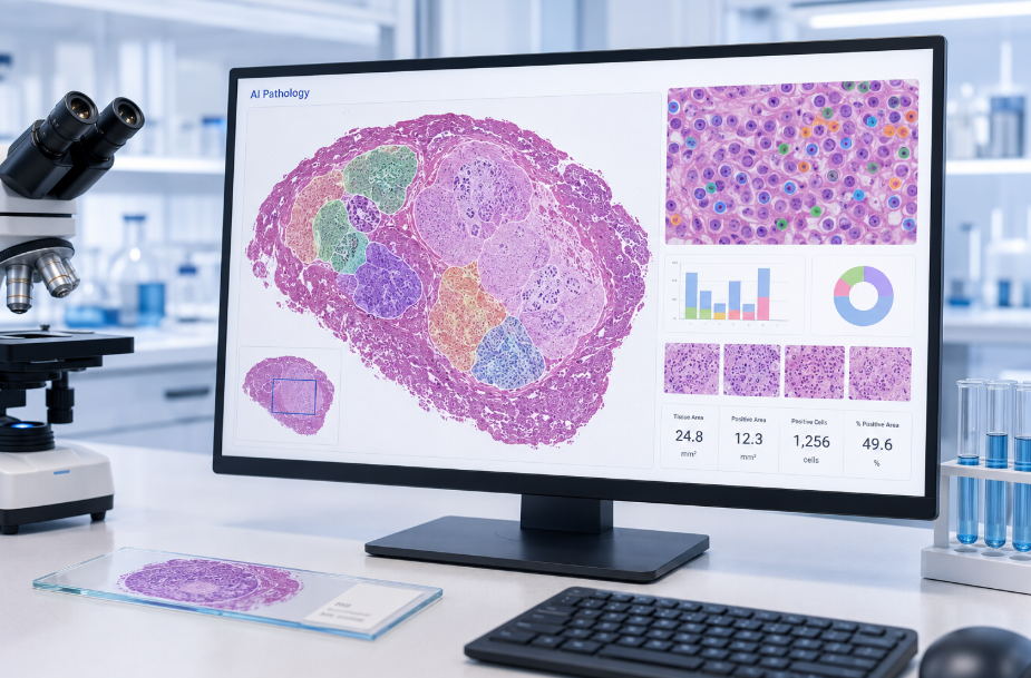
One of the most direct applications is lesion burden measurement. AI models can be trained or configured to identify affected tissue regions, separate them from normal background, and calculate percentage area, total area, regional distribution, and severity-linked image features. In oncology models, this may include tumor area, necrotic fraction, viable tumor compartment, stromal content, or invasive margins. In inflammatory or fibrotic disease models, it may include inflamed tissue area, collagen deposition, epithelial disruption, or tissue remodeling.
These measurements help reduce dependence on broad visual categories and provide continuous variables for statistical analysis. This can be particularly useful in dose-response studies, efficacy screening, and model characterization.
Cell Detection, Classification, and Density Mapping
Cell-level quantification is another important application of AI pathology image analysis. Deep learning models can help detect nuclei, classify cell types, measure cell density, and map spatial distribution. Depending on staining strategy and study objective, this may support quantification of tumor cells, immune cells, stromal cells, hepatocytes, epithelial cells, glial cells, or other relevant populations.
In immuno-oncology and inflammation studies, spatial context is often as important as cell count. AI-assisted WSI analysis can help determine whether immune cells are excluded, infiltrating, clustered, or associated with specific tissue compartments. These spatial endpoints can support mechanism-of-action interpretation and help connect pathology findings with molecular or functional readouts.
Biomarker Scoring and Signal Localization
For immunohistochemistry, immunofluorescence, or multiplex-stained slides, AI pathology can support biomarker quantification at both tissue and cellular levels. Common outputs may include positive cell percentage, staining intensity, H-score-like metrics, regional biomarker distribution, co-localization patterns, and compartment-specific expression.
This is valuable when a therapeutic candidate is expected to modulate a pathway, reduce inflammatory signaling, alter immune infiltration, or change disease-associated marker expression. Instead of relying only on representative images, researchers can generate measurable biomarker endpoints across the full slide or selected regions of interest.
Key Components of an AI Pathology WSI Workflow
A reliable AI pathology workflow should be designed as a controlled analytical process rather than a simple software run. The quality of the endpoint depends on the quality of tissue preparation, scanning, data organization, model selection, annotation strategy, quality control, and biological interpretation.
1. Study Objective and Endpoint Definition
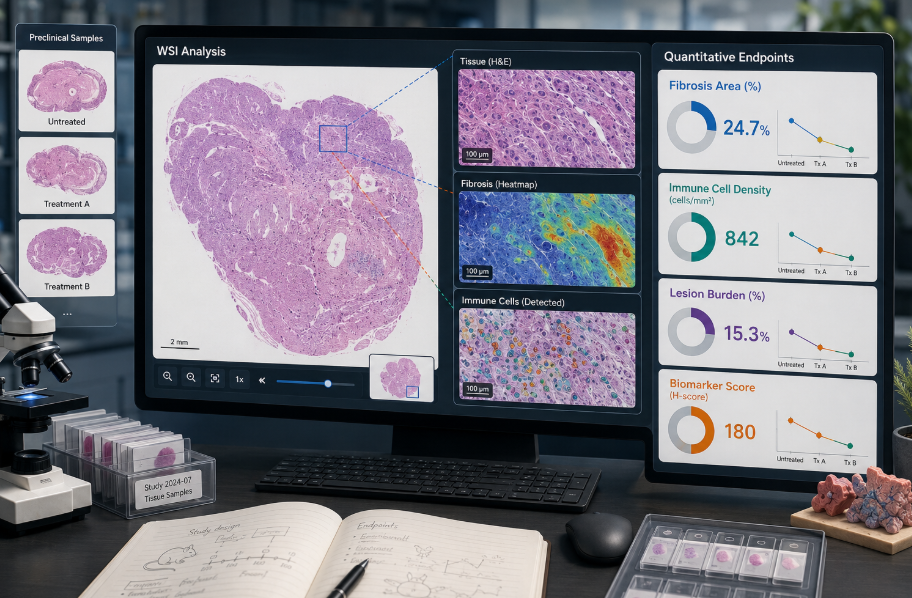
The workflow should begin with the biological question. For example, the goal may be to quantify fibrosis reduction in a liver disease model, measure tumor necrosis after treatment, evaluate inflammatory infiltration in a gut disease model, or assess tissue injury in a toxicology study. Clear endpoint definition helps determine the required tissue type, stain, annotation plan, model architecture, and reporting format.
Poorly defined goals often lead to outputs that are technically impressive but biologically weak. A strong AI pathology workflow defines what should be measured, why it matters, and how the result will be used in preclinical decision-making.
2. Slide Scanning and Image Quality Control
WSI files are large, information-rich, and sensitive to pre-analytical variation. Tissue folds, out-of-focus regions, staining variability, debris, section thickness differences, and scanning artifacts can affect downstream analysis. Therefore, image quality control is essential before model training or batch analysis.
QC should include tissue detection, blur assessment, stain consistency review, artifact exclusion, and confirmation that image resolution is suitable for the target feature. A model designed for tissue-level segmentation may tolerate lower magnification than a model intended for nuclear-level cell classification.
3. Annotation and Ground Truth Development
AI models require well-defined examples of the features they are expected to detect. Depending on the project, annotation may involve region-level labels, pixel-level segmentation, cell-level labels, or slide-level classifications. In preclinical research, annotation quality is especially important because animal models may show tissue patterns that differ from human clinical samples.
Pathologist input remains critical. AI should not replace expert interpretation; it should scale and standardize measurement once the target feature is clearly defined. The FDA’s PathologAI initiative, for example, specifically focuses on AI frameworks for animal-pathology data in preclinical settings, including lesion type, severity, and location analysis in whole-slide images.
4. Model Selection, Training, and Validation
AI pathology pipelines may use classical image analysis, convolutional neural networks, transformer-based models, multiple instance learning, or hybrid approaches. The right method depends on whether the endpoint is local or global, supervised or weakly supervised, cell-based or tissue-based, and stain-specific or stain-agnostic.
Validation should be built into the workflow. This may include comparison with expert annotations, cross-validation across batches, evaluation on independent slides, sensitivity to staining variability, and review of failure cases. Systematic reviews of AI in digital pathology continue to emphasize that performance validation is essential before models are applied to decision-making workflows.
5. Batch Analysis and Endpoint Reporting
Once validated for the study context, the AI model can be applied across the WSI dataset. Outputs should be organized in a way that supports statistical analysis and biological interpretation. A useful report may include slide-level summaries, region-level heatmaps, representative annotated images, endpoint tables, QC flags, model confidence indicators, and method descriptions.
The most valuable deliverable is not just an image overlay. It is a decision-ready endpoint package that allows researchers to compare treatment groups, identify trends, correlate tissue findings with other data layers, and determine whether a candidate or model is worth advancing.
Practical Endpoints Generated from AI Pathology Image Analysis
AI pathology and WSI analysis can support many types of preclinical endpoints, including:
- Tumor area, necrosis ratio, invasive margin features, and stromal proportion
- Fibrosis area, collagen distribution, and tissue remodeling indices
- Inflammatory cell density, immune infiltration patterns, and regional clustering
- Tissue injury area, degeneration scores, and organ-specific lesion burden
- Biomarker positivity, staining intensity, and compartment-specific expression
- Cell density, nuclear morphology, mitotic activity, and spatial organization
- Disease model severity metrics and treatment-response indicators
These endpoints can be used in efficacy studies, toxicity evaluation, pharmacodynamic assessment, disease model validation, antibody screening support, and mechanism-of-action studies.
How AI Pathology Supports Preclinical Decision-Making
A well-designed WSI analysis workflow can improve preclinical research in several ways.
First, it increases measurement consistency. When a validated model is applied across all slides under controlled conditions, the same feature definitions are used throughout the dataset. This can reduce variability caused by manual scoring differences.
Second, it improves scalability. Large preclinical studies may involve hundreds or thousands of slides. AI-assisted analysis can process high-volume datasets more efficiently, enabling broader tissue coverage and more detailed feature extraction.
Third, it enables deeper phenotyping. AI can quantify subtle morphological patterns that may be difficult to capture using traditional categorical scoring alone. This is particularly useful in complex disease models where treatment effects may be spatial, heterogeneous, or pathway-specific.
Fourth, it strengthens data integration. Quantitative pathology endpoints can be correlated with molecular data, pharmacokinetics, pharmacodynamics, clinical chemistry, flow cytometry, cytokine panels, or behavioral readouts. This helps build a more complete picture of candidate activity and model relevance.
Challenges to Consider Before Starting an AI Pathology Project
AI pathology is powerful, but it is not automatic. Several challenges should be addressed early.
Data Quality and Standardization
Model performance depends heavily on image quality, staining consistency, tissue preparation, and metadata accuracy. Batch effects can produce false signals if not controlled. Standardized slide preparation and scanning protocols are therefore important for reproducible WSI analysis.
Biological Specificity
A model trained for one tissue, species, stain, disease model, or endpoint may not perform well in another context. Preclinical studies often use specialized animal models, engineered disease systems, or treatment-induced phenotypes. Customization may be necessary when existing models do not match the study design.
Interpretability and Review
Researchers need to understand what the model is measuring and where it may fail. Heatmaps, overlays, region-level outputs, and expert review are important for interpretability. The best workflows combine computational efficiency with pathology oversight.
Statistical Planning
Quantitative endpoints should be planned with downstream statistics in mind. Study teams should consider sample size, biological replicates, region selection, batch structure, and how slide-level or animal-level measurements will be aggregated.
Best Practices for Turning WSI into Reliable Preclinical Endpoints
To maximize the value of AI pathology image analysis, preclinical teams should define endpoints before slide scanning, standardize tissue and staining procedures, include representative training and validation samples, involve pathology experts in annotation and review, document QC criteria, and report both quantitative values and visual evidence.
The strongest workflows are iterative. Initial model outputs should be reviewed, refined, and validated before final batch analysis. When AI outputs align with biological expectations and expert interpretation, they can become highly useful endpoints for candidate ranking, model comparison, and translational planning.
Conclusion
AI pathology image analysis is transforming WSI from a static digital representation of tissue into a source of quantitative, reproducible, and biologically meaningful endpoints. For preclinical studies, this shift can improve how researchers evaluate efficacy, safety, disease model performance, biomarker modulation, and tissue-level mechanism of action.
The goal is not to replace expert pathology, but to extend it. By combining whole-slide imaging, AI-based feature extraction, quality control, and expert interpretation, research teams can generate pathology endpoints that are more scalable, comparable, and decision-ready. As preclinical programs become increasingly data-driven, AI pathology and WSI analysis will play a growing role in connecting tissue morphology with therapeutic strategy.
Related AI Services from Creative Biolabs
Creative Biolabs provides AI-supported solutions for preclinical research and pathology image analysis. Relevant services include:
- AI-Driven Pathology Image Analysis Service — WSI analysis, pathological feature detection, quantitative reporting, and annotated image deliverables.
- AI-Driven Preclinical Research Service — AI-enabled preclinical data analysis, predictive modeling, and wet-lab-supported research workflows.
- AI-Driven Disease Model Construction & Prediction Service — disease model prediction and data-driven model construction support.
- AI-Driven Antibody Screening Service — AI-assisted antibody screening support for candidate prioritization and downstream research.